Superficial Temporal Artery to Middle Cerebral Artery Anastomosis

Steven D. Chang, M.D., and Gary K. Steinberg, M.D., Ph.D.

Department of Neurosurgery and the Stanford Stroke Center, Stanford University Medical Center, Stanford, California, U.S.A


Abstract: Superficial temporal artery to middle cerebral artery revascularization has evolved has evolved as a method to treat selected patients with intracranial ischemia. Specific indications include nonatherosclerotic occlusive vascular disorders, symptomatic brain ischemia in patients with inaccessible atherosclerotic occlusive disease who have not responded to maximal medical therapy, carotid artery dissection or penetrating injuries, and as an adjunct for deliberate large vessel arterial occlusion after failure of temporary trial occlusion. Careful preoperative evaluation and meticulous attention to detail before surgery yield good patient outcomes with minimal adverse effects and few deaths. This study reviews the indications and operative techniques for superficial temporal artery to middle cerebral artery anastomosis, the most common extracranial to intracranial revascularization procedure.


Superficial temporal artery to middle cerebral artery (STA-MCA) bypass surgery was developed as a treatment for patients with ischemic cerebrovascular disease secondary to vascular lesions that are inaccessible by carotid endarterectomy. Direct measurements of blood pressure within middle cerebral artery branches showed significantly decreased pressures in patients with intracranial occlusive disease who experienced transient ischemic attacks (TIAs) or minor stroke. A direct bypass to the middle cerebral artery would provide a collateral channel for additional blood flow to these ischemic areas and, in theory, improve perfusion.


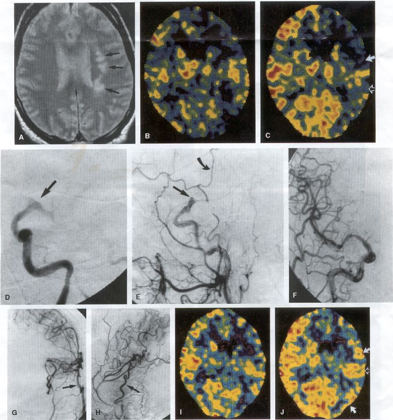
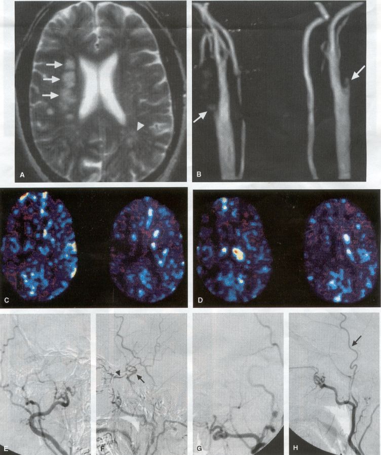
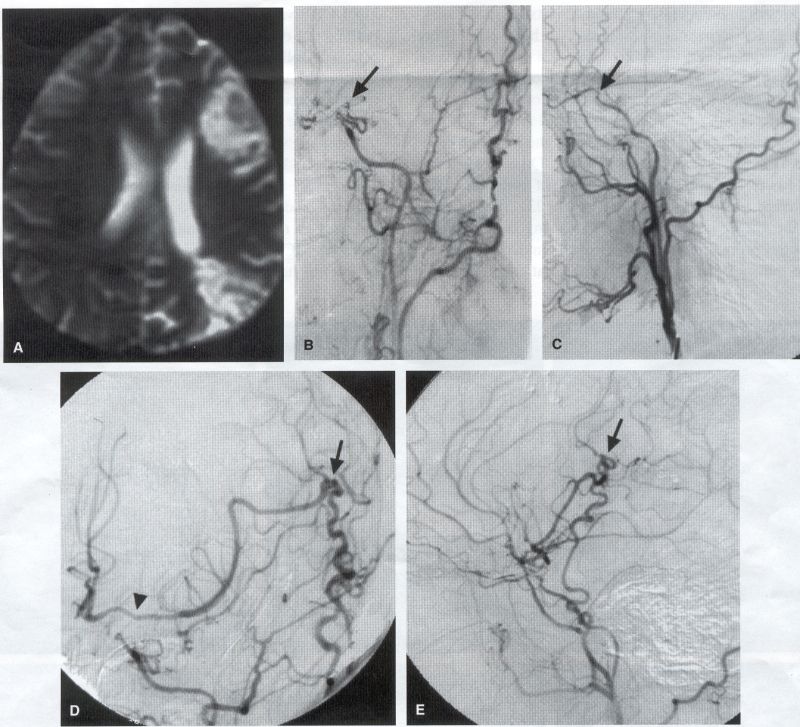
COMMON INDICATIONS FOR SUPERFICIAL TEMPORAL ARTERY TO MIDDLE CEREBRAL ARTERY BYPASS

Atherosclerotic Disease

The most common indication for STA-MCA bypass has been symptomatic atherosclerotic disease of the intracranial internal carotid artery (Figs. 1, 2, 3) or the MCA (Figs. 3, 4). These patients experience decreases in cerebral blood flow (CBF) that produce what has been called "misery perfusion." Ischemic regions of cortex could benefit from collateral flow and should decrease patient symptoms related to low CBF. The most frequently performed STA-MCA bypass is a direct arterial anastomosis from a branch of the STA to an MCA branch emerging from the sylvian fissure. Such grafts have been shown to result in additional blood flow of 25 to 50 mL/min, which may increase as the graft matures (9). Increased flow rates may be obtained with interposition vein grafts, which deliver flows of as much as l00 mL/min immediately after anastomosis, or by grafting the STA to more proximal M2 segments within the sylvian fissure (9,57). Yasargil et al.(67) performed the first successful microvascular STA in 1967. Subsequent studies show that the procedure had high long-term patency rates and low perioperative morbidity and mortality rates (6,7,9,11,15,44,45,58). Two of these studies documented improved regional CBF and anecdotal improvement in clinical symptoms and decreased risk for risk for future stroke (59,68). These series also revealed patency rates ranging from 87% to 100%; mortality rates of 0% to 4.4%; and low morbidity rates for strokes (2.7%), TIAs (7.3%), seizures (5.4%), hemorrhages (4%), and wound infection (1%) (6,7,9,11,15,44,45,59,68). However, these series lacked a matched control population treated with medical therapy and followed various methods of patient selection and evaluation criteria. In an attempt to clarify this issue, the International Cooperative Extracranial-lntracranial bypass study was begun in 1977 and completed in 1985 (1). This study failed to show a benefit of surgical bypass over medical management with aspirin in reducing the risk for fatal or nonfatal strokes, and it resulted in further questions regarding patient selection. The study was criticized for the large number of patients who underwent surgery outside of the study (56), the failure to separate hemodynamic causes of vascular insufficiency from embolic causes (38), the variability in obtaining routine preoperative angiograms and CBF studies (38), and the lack of a trial of medical therapy in many patients before surgical bypass (38).

Recent analysis since the completion of the EC-IC cooperative study seems to show that a select group of patients may benefit from STA-MCA bypass procedures. These patients have CBF and metabolism studies, such as xenon computed tomography or positron emission tomography scans, that indicate poor or absent vascular reserve (18,19,26,33,40,47,65), and they have not responded to medical management with recurrent symptoms while taking antiplatelet agents or anticoagulation. Generally their angiograms show poor collateral flow and often an occluded ipsilateral carotid artery. Two studies have shown that patients with carotid stenosis or occlusion and impaired hemodynamic reserve have a 12-fold greater risk for stroke during a 2-year period compared with similar patients with carotid stenosis or occlusion but without impaired reserve (36% stroke rate versus 4% stroke rate) (63). Other studies confirm a significantly higher ipsilateral stroke rate for patients with carotid occlusion with compromised hemodynamic reserve (16,25,26,66). Patients with carotid occlusion and impaired hemodynamic reserve have a 6.4% to 14% annual ipsilateral stroke rate, whereas patients with severely impaired hemodynamic measurements have a 16.2% to 59.7% annual ipsilateral stroke rate (25,26,41,63,66,70). By comparison, all symptomatic patients with carotid occlusion (without CBF measurements) have an annual ipsilateral stroke rate of 1.6% to 2.8% (25,26,41,63,66,70). Usually no other alternative treatments are available to such patients with impaired reserve. With careful preoperative evaluation, they are the most likely to benefit from STA-MCA bypass. Gewirtz et al. (13) reviewed the Stanford series of 14 patients who did not respond to medical therapy after a carotid artery occlusion and who had poor hemodynamic reserve (with paradoxical steal) based on acetazolamide xenon computed tomographic scans. Thirteen of the patients had no further strokes at a mean follow-up of 49 months (range, 8-77 months). One patient (7%) experienced a stroke 2 weeks after his STA-MCA procedure when he underwent emergent coronary artery bypass surgery for unstable angina and became hypotensive during surgery. Of the nine patients who underwent a second xenon computed tomographic scan after surgery, eight patients showed improved hemodynamic reserve after acetazolamide therapy. Other investigators also found favorable clinical results after STA-MCA bypass in patients with internal carotid artery occlusion and impaired hemodynamic reserve (47.62).

Moyamoya Disease

Moyamoya disease is a progressive disease of the internal carotid artery, MCA, and anterior cerebral artery characterized by occlusion of these vessels, which show intimal proliferation (2,17,36,64). Children with this disease usually have strokes, whereas adults have ischemic symptoms or intracranial hemorrhage. The natural history of untreated moyamoya disease is poor, with a 73% rate of major deficit or death more than 2 years after diagnosis in children (36) and a similarly poor prognosis in adults (24,43). The first STA-MCA bypass for moyamoya disease was performed by Yasargil and Yonekawa in 1972 (68), and since then several small series have been reported. Karasawa et al. described 12 patients with moyamoya disease who underwent bypass, with 10 patients (83%) having good to excellent results (24). Quest and Correll reported a similar outcome in 11 of 13 patients (85%) (43). Ishikawa et al. (22) compared STA-MCA bypass (48 procedures) with indirect revascularization methods (16 procedures), such as encephalo-duro-arterio-myosynangiosis, in pediatric patients with moyamoya disease and found that the incidence of postoperative ischemic events was significantly decreased in the direct bypass group (10%) compared with the indirect group (56%; p <0.01). They concluded that direct revascularization is the procedure of choice over indirect revascularization whenever possible. Matsushima et al. (31) found a similar advantage of direct revascularization compared with indirect methods, although with a smaller group of patients. At Stanford, since 1991 we have performed 59 (as of June 2000) STA-MCA grafts in patients with moyamoya disease (ages 5 to 63; Fig. 4) with excellent overall clinical results (5,14,55). Six patients had temporary minor worsening after operation that resolved with volume expansion, and one patient had a postoperative hemorrhage into a previously ischemic region. The mean follow-up was 43 months, ranging from 1 month to 7 years, and 95% of patients were neurologically stable or improved. Persistent TIAs developed in one patient after occlusion of a STA-MCA bypass with short vein interposition. Follow-up angiography showed that 97% of grafts were patent, and hemodynamic reserve was shown to be improved on CBF studies in most patients. There were no perioperative deaths, but one adult died of a myocardial infarction 72 months after bypass, and another adult died 18 months after an external carotid to MCA bypass using a vein interposition (from hemorrhage into ischemic brain). Of 12 children with moyamoya disease (in 19 hemispheres) treated with STA-MCA bypasses (8 after strokes and in 5 with refractory TIAs), none have experienced new strokes (mean follow-up, 35 months; range, 12-65 months) (14). Postoperative angiograms show successful revascularization of the MCA distribution in 89% of the cases, and postoperative xenon computed tomographic scans showed increased augmentation with acetazolamide compared with the results of the preoperative study (14).


PREOPERATIVE RADIOGRAPHIC EVALUATION

Bilateral carotid angiograms are performed to evaluate donor and recipient vessels and collateral circulation. The size and location of optimal vessels to be used for the anastomosis are identified. A balloon test occlusion can be performed to determine the tolerance of particular vessels if planned occlusion is required. Patient who have poor results of this test need prophylactic revascularization. Xenon computed tomographic CBF studies (Figs. 1, 2, 3) or provocative induced hypotension during test occlusion may provide additional information regarding tolerance to occlusion (50,54,69). Intraoperative electroencephalography, microvascular Doppler studies, and somatosensory evoked potentials are useful adjuncts. Postoperative bypass patency is usually evaluated with conventional cerebral angiography. but recent studies show that the results of magnetic resonance angiography correlate reasonably well with those of conventional angiography (27,28,42)


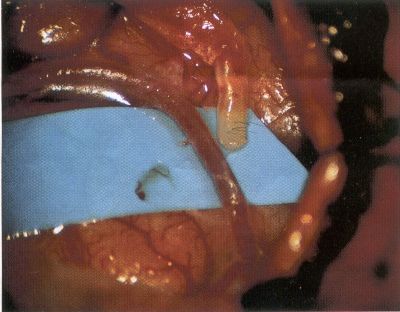
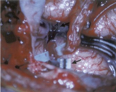
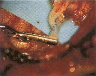
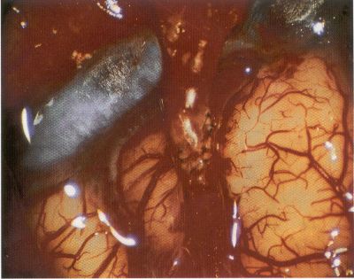
OPERATIVE TECHNIQUE

General Principles

Several operative generalities are applicable to STA-MCA revascularization procedures. The patient is positioned with his or her head above the heart to reduce venous cerebral congestion. Doppler ultrasound is used to identify the location of donor vessels. Hyperventilation and a-adrenergic agents are not recommended because of their vasoconstrictive effects, but mild hypothermia (33°C) and barbiturates are used routinely to provide a cerebral protective effect during arterial occlusion. Vasoconstrictive agents such as epinephrine should not be used during scalp infiltration. Intraoperative monitoring with electroencephalography and evoked potentials provide early detection of ischemic changes.

Microinstruments and the operating neurosurgical microscope are used routinely for revascularization procedures. Donor vessels should be selected with a diameter ³1 mm because smaller vessels have higher occlusion rates, provide less blood flow, and are more difficult to anastomose. Short vein interposition grafts between the proximal STA and MCA branches may be used when the distal MCA is too small or not available. Papaverine is useful in preventing vascular spasm.

If a short venous interposition graft is needed, the saphenous vein is the vessel of choice. It is harvested adjacent to the ankle, side branches are carefully ligated, and care is taken to avoid any laceration to the vein itself. The vein is irrigated with heparinized saline and inspected for any lacerations that would require surgical repair before use. The vein is stored in heparinized saline until it is needed. Sometimes the superficial temporal vein can be used for a short interposition graft if its diameter is sufficiently large.

Hemodynamic control is the primary goal of postoperative management. Hypertension may result in excessive bleeding at the anastomotic site. Intraparenchymal hemorrhages, from increased perfusion of the brain, and a possible leak in the anastomotic site are other complications. Conversely, hypotension may cause graft occlusion, resulting in clinical ischemia. In such cases, an emergent angiogram and graft revision may be necessary. Prophylactic anticoagulants may be started on the first postoperative day, although aspirin is usually adequate. A cerebrospinal fluid leak is a potential complication, particularly when vein grafts are used, because of the deliberately loose dural closure, which prevents excessive pressure on the donor vessel.

Operative Technique for Superficial Temporal Artery to Middle Cerebral Artery Bypass With or Without Short Vein Graft

The patient is positioned with the head turned to the contralateral side and the temporal bone parallel to the floor, and the patient's head is fixed in a three-point Mayfield headrest. After the hair is shaved from the scalp, a Doppler ultrasound is used to mark out the course of the STA and its branches (Fig. 8). After local infiltration of the scalp, an incision is made with a #15 blade and the superficial temporal artery is identified and harvested with a generous cuff of soft tissue surrounding the artery (Fig. 9). Either the frontal or parietal STA branch may be used, depending on diameter size and suitable length. Smaller branches of the artery are isolated and carefully coagulated to prevent injury to the STA or they are tied off. The length of the artery required depends on the distance from the artery to the graft site and on whether a short vein segment will be used. A craniotomy is performed in the midfrontal-temporal bone overlying the sylvian fissure (Fig. 10). The dura is opened and recipient vessels are identified. Optimal recipient vessels have outer luminal diameters ³1 mm. A microvac subdural drain is placed to remove cerebrospinal fluid during operation.

The recipient MCA/branch (usually the M3 or M4 branch emerging from the sylvian fissure) is chosen (Fig. 11) based on diameter, location (the angular MCA branch is the optimal recipient), and orientation (perpendicular to the surgeon if possible). Dissection of the arachnoid layer over a 6- to 10-mm segment exposes this vessel, and tiny branches are coagulated and divided if necessary. A high-visibility background is placed to facilitate anastomosis (Fig. 12). A temporary aneurysm clip is placed on the proximal STA donor, and the distal STA is ligated and divided at the proper length. The temporary clip is opened briefly to verify good flow through the STA. Then the STA is flushed with heparinized saline and the distal tip of this vessel is stripped of all fascial tissue and cut in an oblique manner (Fig. 13). Microvascular clips (Fig. 14) or temporary aneurysm clips (Fig. 15) are placed on each side of the recipient vessel to prevent bleeding. An arteriotomy is performed by cutting an elliptical or diamond-shaped portion of the superior wall of the recipient vessel. The recipient branch is irrigated with heparinized saline. Indigo carmine blue dye can be used to stain the edges of the MCA vessel, thereby facilitating suturing. Anastomosis is performed under the neuromicroscope using 10-0 interrupted monofilament suture (or alternatively, running 10-0 monofilament suture) (Fig. 16). Corner stitches are placed first, followed by stitches at the far wall, and finally those at the near wall. The intimal layer must be included with each interrupted stitch, but significant narrowing of the anastomotic site should be avoided. Temporary clips are removed. Significant bleeding may indicate the need for an additional stitch, whereas small amounts of bleeding may be treated with Gelfoam or Surgicel. If the donor vessel is a short vein segment, a proximal anastomosis to the STA is performed first using 10-0 or 9-0 monofilament suture. The vein must be of an appropriate length to avoid any kinks in the vein. An arteriotomy is made in the superficial temporal artery, and 10-0 monofilament sutures are used to anchor the corner stitches, followed by stitches at the back and the front walls. Once the anastomosis is complete (Figs. 17, 18, 19), a Doppler ultrasound may be used to test patency. We have found a quantitative and directional Doppler useful in the measurement of flow through the MCA segment and the STA before anastomosis, and in the STA and proximal and distal MCA segments after bypass. The dura is then closed loosely around the STA graft and the bone is trimmed to avoid any compromise of the STA. The galeal layer and skin are closed, with care taken to avoid graft compromise.


POSTOPERATIVE ISSUES

Patency, Morbidity, and Mortality Rates

Many large series of STA to MCA bypass grafts are reviewed in the literature. They reveal patency rates ranging from 87% to 100%, mortality rates of 0 to 4.4%, and morbidity rates for strokes (2.7% ), TIAs (7.3% ), seizures (5.4%), hemorrhages (4%), and wound infection (1%) (6,7,9,11,15,44,45,59,68). The EC-IC study noted a patency rate of 96% and a mortality rate of 0.6%.

Long- Term Effects of Revascularization

Several authors have examined the long-term effects of STA-MCA bypass on cerebral circulation. Tsuda et al. (60) showed (using SPECT studies) improved cortical regional CBF and cognition 3 years after the bypass. Similarly, positron emission tomography scans have shown increases in regional CBF and decreases in oxygen extraction fraction after revascularization (32,35).

Complications

There are several potential complications of STA-MCA bypass procedures. Ischemic changes usually result from either compression of the donor vessel by the scalp closure, graft occlusion or stenosis at the anastomosis site, or emboli. Doppler and angiography studies generally identify the cause. As discussed above, hypertension may result in anastomosis leak or parenchymal hemorrhage, and increased perfusion may cause cerebral edema and disautoregulation. In addition, because a loose dural closure is necessary to avoid any compression of the donor vessel, cerebrospinal fluid leak, pseudomeningocele, and subdural hygromas are possible, although rare. Scalp ischemia, although a theoretical risk, is extremely unusual. Finally, as with other craniotomies, systemic surgical complications include myocardial infarction, pneumonia, deep venous thrombosis, and pulmonary emboli.


CONCLUSIONS

Extracranial-to-intracranial (STA-MCA) revascularization procedures have proved useful in selected patients with cerebral ischemia. Judicious preoperative evaluation coupled with attentive intraoperative techniques and postoperative management have minimized morbidity and mortality rates while improving patient outcome. Improvements in our understanding of the indications for such procedures should further refine the patient population that will benefit from these revascularization procedures.


FIG. 1

Figure 1

A 43-year-old man had expressive aphasia and mild right hemiparesis as a result of persistent left hemisphere TIAs despite therapeutic warfarin sodium and low molecular weight heparin. (A) A preoperative magnetic resonance image showed watershed infarct in the left centrum semiovale deep white matter (arrows). Pretreatment xenon computed tomographic imaging (B) before and (C) after acetazolamide administration revealed impaired hemodynamic reserve with paradoxical steal in the left frontoparietal region (arrows). The right hemisphere showed normal augmentation of cerebral blood flow after acetazolamide. A left common carotid cerebral angiogram showed occlusion of the AI anterior cerebral and MI middle cerebral arteries (straight arrow), seen in the (D) anteroposterior and (E) lateral views. The parietal branch of the left superficial temporal artery (E, curved arrow) was later used as the donor vessel for superficial temporal artery to middle cerebral artery (STA-MCA) bypass. (F) A right anteroposterior internal carotid artery cerebral angiogram revealed absence of aright A1 anterior cerebral artery and no filling of the contralateral left middle cerebral artery territory. No posterior communicating arteries were visualized on carotid or vertebrobasilar injections. The patient underwent a left STA-MCA bypass with subsequent resolution of his TIAs. (G) A postoperative anteroposterior and (H) lateral left external carotid cerebral angiogram (7 weeks after surgery) showed marked enlargement of the STA (arrow) with excellent filling of the middle cerebral and anterior cerebral artery territories from the STA graft. A postoperative xenon CT scan (also 7 weeks after surgery) (I) before and (J) after acetazolamide challenge showed improved hemodynamic reserve in the left frontoparietal regions (arrows). After operation, the patient has had no further TIAs or strokes while taking aspirin during the last 5 years.

Back to top

FIG. 2

Figure 2

A 61-year-old man had acute left hemiparesis and a single TIA consisting of right arm weakness. (A) A T2 axial magnetic resonance image of his head shows multiple right hemisphere white matter infarcts (arrows) and a left parietal infarct (arrowhead). (B) A magnetic resonance angiogram of the patient's neck revealed bilateral internal carotid artery occlusions (arrows). A xenon computed tomographic scan was performed and showed (C) poor baseline cerebral blood flow bilaterally and (D) minimal augmentation after the administration of acetazolamide. A cerebral angiogram performed with a right common carotid artery injection showed primarily external carotid vessels in the (E) anteroposterior and (F) lateral views. The right supraclinoid carotid artery (arrow, F) fills slightly via the right ophthalmic artery (arrowhead, F). A left common carotid artery angiogram shows filling of the external carotid branches only, seen on (G) anteroposterior and (H) lateral views (the arrow shows the superficial temporal artery [STA] on the lateral view). The patient underwent aright superficial temporal artery to middle cerebral artery (STA-MCA) bypass procedure, followed 6 weeks later by a left STA-MCA bypass.

Back to top

FIG. 3

Figure 3

Same patient as in Figure 2. Three weeks after the second bypass, a right external carotid angiogram showed excellent filling of the MCA distribution in the (A) anteroposterior and (B) lateral views. The anastomosis site (arrow, A and B) can be seen in both views, as can retrograde flow down the proximal MCA (arrowhead, A). A left external carotid angiogram showed excellent filling of the left MCA in the (C) anteroposterior and (D) lateral views with the anastomosis site visible (arrow. D). The patient's left hemiparesis resolved over 2 months with rehabilitation and he was neurologically normal. He has had no further TIAs.

Back to top

FIG. 4

Figure 4

A 35-year-old woman had right hemiparesis and expressive aphasia after a left cerebral stroke. Evaluation revealed bilateral moyamoya disease. (A) A pretreatment T2 MRI axial image shows watershed distribution infarcts in the left hemisphere. A left common carotid cerebral angiogram revealed poor filling above the supraclinoid internal carotid artery (arrow) in the (B) anteroposterior and (C) lateral views. She underwent a right superficial temporal artery to middle cerebral artery (STA-MCA) bypass, followed 1 week later by a left STA-MCA bypass. Post-treatment angiography showed excellent filling of the MCA vessels bilaterally from the STA-MCA grafts. A left common carotid cerebral angiogram shows the STA-MCA anastomosis site (solid arrow) in both the (D) anteroposterior and (E) lateral views. Retrograde filling down the MCA to the Al-Ml junction (D, arrowhead) was evident, as was filling of the bilateral A2 anterior cerebral arteries from the graft. The patient has had no further ischemic symptoms and mild improvement in her aphasia 1 year after surgery.

Back to Moyamoya Disease


FIG. 8

Figure 8

A typical skin incision. The main incision is planned over the superficial temporal artery (STA), with a T extension to allow exposure of the bone.

Back to Operative Technique

FIG. 9

Figure 9

The initial superficial temporal artery (STA) preparation involves making a linear incision over the surface (A) with careful dissection of the main STA branch (B).

Back to Operative Technique


FIG. 10

Figure 10

After the superficial temporal artery (STA) is dissected out, the temporalis muscle is divided and the bone flap is made to allow exposure of the brain over the anterior sylvian fissure.

Back to Operative Technique


FIG. 11

Figure 11

A middle cerebral artery (MCA) recipient vessel is identified. The sylvian fissure can be split to allow identification of a larger, more proximal branch of the MCA, preferably an M3 branch.

Back to Operative Technique


FIG. 12

Figure 12

The end of the donor superficial temporal artery (STA) adjacent to the recipient MCA (M3) artery. A blue high-visibility backfield is placed behind the middle cerebral artery (MCA) branch to increase suture visibility.

Back to Operative Technique


FIG. 13

Figure 13

The distal tip of the superficial temporal artery (STA) is cut in a fish-mouth manner to facilitate anastomosis. A small scissors is used to remove an oval segment of the middle cerebral artery (MCA) branch.

Back to Operative Technique


FIG. 14

Figure 14

An intraoperative photograph shows occlusion of the MCA branch with temporary microvascular clips (arrows).

Back to Operative Technique


FIG. 15

Figure 15

An intraoperative photograph shows occlusion of the MCA branch with temporary aneurysm clips.

Back to Operative Technique


FIG. 16

Figure 16

After placing vascular clips on both sides of the anastomosis site and preparing both vessels, (A) the two end sutures are placed first, followed by (B) interrupted sutures along the front and back wall of the anastomosis. (C) When the anastomosis is complete, the vascular clips are removed.

Back to Operative Technique


FIG. 17

Figure 17

The completed anastomosis shows the superficial temporal artery (STA) artery positioned such that flow is directed toward the proximal portions of the middle cerebral artery (MCA).

Back to Operative Technique


FIG. 18

Figure 18

Completion of the superficial temporal artery (STA) anastomosis before removal of the high-visibility backfield.

Back to Operative Technique


FIG. 19

Figure 19

The final superficial temporal artery to middle cerebral artery (STA-MCA) anastomosis.

Back to Operative Technique


There are user(s) in the live chat room. Go there now...
Main
Homepage

DJ's Story
Donate
Pictures
Moyamoya Family
Kansas City 2012
Philadelphia 2011
Las Vegas 2008
San Antonio 2007
Orlando 2006
Las Vegas 2005
SPECT Scans

DJ's 1st Surgery
DJ's 2nd Surgery
Medical Info
Doctor Videos!
Moyamoya?
STA-MCA
Surgeries
Communicate
Guest Book
Message Board

Archives
LIVE Chat!
Shop MM.com
Online Store
Links
Links Page

Netiquette


DISCLAIMER: All information contained on this web site is for informational purposes only.  It is in no way intended to be used as a replacement for professional medical treatment. moyamoya.com makes no claims as to the scientific/clinical validity of the information on this site OR to that of the information linked to from this site.  All information taken from the internet should be discussed with a medical professional!


Click here to email DJ


©2003-2018 Web Vision Enterprises LLC. All rights reserved. All information on this site is protected by international
copyright laws. You may not re-distribute any information from this site without written permission
from
Web Vision Enterprises LLC and the webmaster of this site. Violators will be prosecuted.
You may view our privacy policy and financial disclosure statement here


REFERENCES

1. Failure of extracranial-intracranial arterial bypass to reduce the risk of ischemic stroke. Results of an international randomized trial. The EC/IC Bypass Study Group. N Engl J Med 1985;313:1191- 1200.
2. Carlson CB, Harvey FH, Loop J. Progressive alternating hemiplegia in early childhood and basal arterial stenosis and telangiectasia (moyamoya syndrome). Neurology 1973;23:734-44.
3. Chang SD, Steinberg GK. Other treatment for stroke prevention. In: Bogousslavsky J, Ginsberg M, eds. Cerebrovascular disease. London: Blackwell Scientific Publications, 1998:1945-63.
4. Chang SD, Steinberg GK. STA-MCA: Its use and prediction of outcome. In: Fisher WS, ed. Perspectives in neurological surgery. Volume 9. Baltimore: Williams and Wilkins. 1998:43-61.
5. Chang SD, Steinberg GK. Surgical management of moyamoya disease. In: Tindall GT, ed. Contemporary neurosurgery. Volume 22, No.10. Baltimore: Lippincott Williams and Wilkins, 2000:1-10.
6. Chater N. Neurosurgical extrilcraniaI-intracranial bypass for stroke: with 400 cases. Neurol Res 1983;5:1-9.
7. Collice M, Arean 0, Eermino F, Riva M. Surgical and long term results in 100 consecutive patients with ICA occlusion. In: Spetzler RF, Carter LP, Selman WR, Martin N, eds. Cerebral revascular- izationfor stroke. New York: Thieme-Stratton, 1985:445-9.
8. D' Alise MD, Vardiman AB, Kopitnik TA Ir, Batjer HH. External carotid-to-middle cerebral bypass in the treatment of complex in- ternal carotid injury. J Trauma 1996;40:452-5.
9. Eguchi T. Results of EC-IC bypass with and without long vein graft. In: Spetzler RF, Carter LP, Selman WR, Martin N, eds. Cerebral revascularization for stroke. New York: Thieme- Stratton, 1985:584-90.
10. Eguchi T, Mayanagi Y, Hanamura T, Tanaka H, Ohmori K, Takakura K. Treatment of bilateral spontaneous carotid-cavernous fistula by Hamby's method combined with an extracranial- intracranial bypass procedure. Neurosurgery 1982; II :706-11.
11. Gagliardi R, Benvenuti L, Onesti S. Seven years experience with extracranial-intracranial arterial bypass for cerebral ischemia. In: Spetzler RF, Carter LP, Selman WR, Martin N, eds. Cerebral revascularization for stroke. New York: Thieme-Stratton, 1985:413-25.
12. Gewertz BL, Samson DS, Ditmore QM, Bone GE. Management of penetrating injuries of the internal carotid artery at the base of the skull utilizing extracranial-intracranial bypass. J Trauma 1980;20: 365-9.
13. Gewirtz RI, Marks MP, Steinberg GK. Clinical outcome of symp- tomatic patients with carotid artery occlusion and decreased he- modynamic reserve treated with STA-MCA bypass [abstract]. J Neurosurg 1997;86:352A.
14. Golby AJ, Marks MP, Thompson RC, Steinberg GK. Direct and combined revascularization in pediatric moyamoya disease. Neu- rosurgery 1999;45:50-60.
15. Gratzl 0, Roun I. Quality grading of bypass surgery. Operative combined risks less than 4 percent. In: Spetzler RF, Carter LP, Selman WR, Martin N, eds. Cerebral revascularizationfor stroke. New York: Thieme-Stratton, 1985:467.
16. Grubb RL, Derdeyn CP, Fritsch SM, Carpenter DA, Yundt KD, Videen TO, Spitznagel EL, Powers WI. Importance of hemody- namic factors in the prognosis of symptomatic carotid occlusion. JAMA 1998;280:1055-60.
17. Halonen If, Halonen V, Donner M, Iivanainen M, Vuolio M, Makinen I. Occlusive disease of intracranial main arteries with collateral networks in children. Neuropadiatrie 1973;4:187-206.
18. Halsey IH Ir, Morawetz RB, Blauenstein OW. The hemodynamic effect of STA-MCA bypass. Stroke 1982;13:163-7.
19. Holzschuh M, Brawanski A, Ullrich W, Meixensberger I. Cerebral blood flow and cerebrovascular reserve 5 years after EC-IC by- pass. Neurosurg Rev 1991;14:275-8.
20. Howard GF, Ho SU, Kim KS:Wallach I. Bilateral carotid artery occlusion resulting from giant cell arteritis. Ann NeuroI1984;15: 204-7.
21. Hunt JL, Snyder WH. Late false aneurysm of the carotid artery: repair with extra-intracranial arterial bypass. J Trauma 1979;19: 198-200.
22. Ishikawa T, Houkin K, Kamiyama H, Abe H. Effects of surgical revascularizationon outcome of patients with pediatric moyamoya disease. Stroke 1997;28:1170-3.
23. Ieffreys RV, Holmes AE. Common carotid ligation for the treat- ment of ruptured posterior communicating aneurysms. J Neurol Neurosurg Psychiatry 1971;34:576-9.
24. Karasawa I. Kikuchi H, Furuse S, Kawamura I, Sakaki T. Treatment of moyamoya disease with STA-MCA an1lstomosis.J Neu- rosurg 1978;49:679-88.
25. Kleiser B. Widder B. Course of carotid artery occlusion with im- paired cerebrovascular reactivity. Stroke 1992;23:171-4.
26. Klijn CJM, KappelleJtI, Tulteken CAF,Qijn.J. Symptomatic ca rotid artery occlusion: A reappraisal of hemodynamic factors. Stroke 1997;28:2084--93.
27. Kodoma T, Suzuki Y, Yano T, Watanabe K, Veda T, Asada K. Phase-contrast MRA in the evaluation of EC-IC bypass patency. Clin Radiol1995;50:459-65.
28. Kodama T, Veda T, Suzuki.Y, Yano T, Watanabe K. MRA in the evaluation of EC-IC bypass patency. J Comput Assist Tomogr 1993;17:922-6.
29. Lawton MT, Hamilton MG, Beals SP, Ioganic EF, Spetzler RF. Radical resection of anterior skull base tumors. Clin Neurosurg 1995;42:43-70.
30. Martin NA, Weinstein PR. Extracranial-intracranial bypass for ischemic related to carotid siphon stenosis after radiation therapy for pituitary tumor. Proceedings of the 7th International Sympo- sium for Microvascular Anastomosis [abstract]. 1984.
31. Matsushima T, Inoue T, Suzuki SO, Fujii K, Fukui M, Hasuo K. Surgical treatment of moyamoya disease in pediatric patients- comparison between the results of indirect and direct revascular- ization procedures. Neurosurgery 1992;31:401-5.
32. Muraishi K, Kameyama M, Sato K, et al. Cerebral circulatory and metabolic changes following EC/IC bypass surgery in cerebral occlusive diseases. Neurol Res 1993;15:97-103.
33. Nagata S, Fujii K, Matsushima T, Fukui M, Sadoshima S, Ku- wabara Y, Abe H. Evaluation of EC-IC bypass for patients with atherosclerotic occlusive cerebrovascular disease: clinical and pos- itron emission tomographic studies. Neurol Res 1991;13:209-16.
34. Nishioka H. Results of the treatment of intracranial aneurysms by occlusion of the carotid artery in the neck. J Neurosurg 1966;25: 660-704.
35. Ogawa A, Kameyama M, Muraishi K, Yoshimoto T, Ito M, Saku- rai Y. Cerebral blood flow and metabolism following superficial temporal artery to superior cerebellar artery bypass for vertebro- basilar occlusive disease. J Neurosurg 1992;76:955-60.
36. Olds MV, Griebel RW, Hoffman HI, Craven M, Chuang S, Schutz H. The surgical treatment of childhood moyamoya disease. J Neu- rosurg 1987;66:675-80.
37. Olteanu-Nerbe V, Marguth F.Extracranial-intracranial bypass op- eration in basal tumors. Neurosurg Rev 1982;5:99-105.
38. Onesti ST, Solomon RA, Quest DO. Cerebral revascularization: a review. Neurosurgery 1989;25:618-28; discussion 628-29.
39. Peerless SI, Hampf CR. Extracranial to intracranial bypass in the treatment of aneurysms. Clin Neurosurg 1985;32: 114--54.
40. Piepgras A, Leinsinger G, Kirsch CM, Schmiedek P. STA-MCA bypass in bilateral carotid artery occlusion: clinical results and long-term effect on cerebrovascular reserve capacity. Neurol Res 1994;16:104--7.
41. Powers WI, Tempel LW, Grubb RL. Influence of cerebral hemo- dynamics on stroke risk: ol\e year follow-up of 30 medically treated patients. Ann NeuroH989;25:325-30.
42. Praharaj SS, Coulthard A, Gholkar A, English P, Mendelow AD. Magnetic resonance angiographic assessment after extracranial- intracranial bypass surgery. JNeurol Neurosurg Psychiatry 1996; 60:439-41.
43. Quest DO, Correll JW. Basal arterial occlusive disease. Neurosur- gery1985;17:937-41.
44. Reale F, Benericetti E, Benvenuti L, et al. Extra-intracranial arte- rial bypass in typical carotid reversible ischaemic deficits: long- term follow-up in 100 patients. Neurol Res 1984;6:113-4.
45. Reichman OH. Complications of cerebral revascularization. Clin Neurosurg 1976;23:318-35.
46. Samson DS. Cervical carotid injuries. Clin Neurosurg 1982;29: 647-56.
47. Schmiedek P, Piepgras A, Leinsinger G, Kirsch CM, Einhupl K. Improvement of cerebrovascular reserve capacity by EC-IC arterial bypass surgery in patients with ICA occlusion and hemodynamic cerebral ischemia. J Neurosurg 1994;81:236-44.
48. Sengupta RP. Special indications of external/internal bypass op- eration. Neurosurg Rev 1982;5:107-12.
49. S~effert GF, Weinstein PR, Moore WS. Bilateral extracranial- intracranial bypass before carotid ligation for hemorrhage into a pharyngocutaneous fistula. Surgery 1982;92:553-60..
50; Smith HA, Thompson-Dobkin J, Yonas H:, Flint E. Correlation of xenon-enhanced computed tomography-defined cerebral blood flow reactivity and collateral flow patterns. Stroke 1994;25: 1784-7..
51. Spetzler RF, Carter LP. Revascularization and aneurysm surgery: current status. Neurosurgery 1985; 16: 111-6..
52. Spetzler RF, Owen MP. Extracranial-intracranial arterial bypass to a single branch of the middle cerebral artery in the management of a traumatic aneurysm. Neurosurgery 1979;4:334-7..
53. Spetzler RF, Selman W, Carter LP. Elective EC-IC bypass for unclippable intracranial aneurysms. Neurol Res 1984;6:64-8..
54. Standard SC, Ahuja A, Guterman LR, et al. Balloon test occlusion of the internal carotid artery with hypotensive challenge. Am J Neuroradiol1995;16:1453-8..
55. Stoodley M, Marks MP, Steinberg GK. Cerebral revascularization for adult moyamoya disease [abstract]. J Neurosurg 1999;90:200A..
56. Sundt TM Jr. Was the international randomizw trial of extracra- nial-intracranialarterial bypass representative of the population at risk? N Engl J Med 1987;316:814-6..
57. Sundt TM Jr, Piepgras DG, Marsh WR, Fode NC. Saphenous vein bypass grafts for giant aneurysms and intracranial occlusive dis- ease. J Neurosurg 1986;65:439-50..
58. Sundt TM Jr, Siekert RG, Piepgras DG, Sharbrough FW, Rouser OW. Bypass surgery for vascular disease of the carotid system. Mayo Clin Proc 1976;51:677-92..
59. Sundt TM Jr, Whisnant JP, Fode NC, Piepgras DG, Rouser OW. Results, complications, and follow-up of 415 bypass operations for occlusive disease of the carotid system. Mayo Clin Proc 1985;60: 230-40..
60. Tsuda Y, Yamada K, Hayakawa T, Ayada Y, Kawasaki S, Matsuo H. Cortical blood flow and cognition after extracranial-intracranial bypass in a patient with severe carotid occlusive lesions. A three- year follow-up study. Acta Neurochir (Wien) 1994;129:198-204..
61. Tsuji N, Kuriyama T, Iwamoto M, Shizuki K. Moyamoya disease associated with craniopharyngioma. Surg Neurol1984;21:588-92..
62. Vorstrup S, Haase J, Waldemar G, Andersen A, Schmidt J, Paulson OB. EC-IC bypass in patients with chronic hemodynamic insuffi- ciency. Acta Neurol Scand SuppI1996;166:79-81..
63. Webster MW, Makaroun MS, Steed DL, Smith HA, Johnson DW, Yonas H. Compromised cerebral blood flow reactivity is a predic- tor of stroke in patients with symptomatic carotid artery occlusive disease. J Vascular Surg 1995;21:338-44..
64. Yamashita M, Oka K, Tanaka K. Histopathology of the brain vas- cular network in moyamoya disease. Stroke 1983;14:50-8..
65. Yamashita T, Kashiwagi S, Nakano S, et al. The effect of EC-IC bypass surgery on resting cerebral blood flow and cerebrovascular reserve capacity studied with stable XE-CT and acetazolamide test. Neuroradiology 1991;33:217-22..
66. Yamauchi H, Fukuyama H, Nagahama Y, et al. Evidence for mis- ery perfusion and risk for recurrent stroke in major cerebral arterial occlusive diseases from PET. J Neurol Neurosurg Psychiatry 1996;61:18-25..
67. Yasargil MG, Krayenbuhl HA, Jacobson JR. Microneurosurgical arterial reconstruction. Surgery 1970;67:221-33..
68. Yasargil MG, Yonekawa Y. Results of microsurgical extra- intracranial arterial bypass in the treatment of cerebral ischemia. Neurosurgery 1977;1:22-4..
69. Yonas H, Jungreis C. Xenon CT cerebral blood flow: past, present, and future [letter; comment]. Am J NeuroradioI1995;16:219-20..
70. Yonas H, Smith HA, Durham SR, Pentheny SL, Johnson DW. Increased stroke risk predicted by compromised cerebral blood flow reactivity. J Neurosurg 1993;79:483-9.